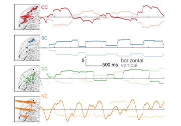
A new paper by José Ossandón, University of Hamburg, Germany, Dr Ramesh Kekunnaya, the Child Sight Institute at LVPEI, and others, systematically investigates active visual exploration behavior among those with restored vision from congenital blindness. They present evidence that eye movements necessary for object perception emerge even if sight is restored after extended periods of congenital blindness.
We move our eyes and systematically scan our environment to visually perceive the world. Research has shown that our eye movements are guided by low-level features, like luminance contrast and high-level features, like object components. Eye movements are crucial for object recognition which requires the integration of multiple features like size, color, surface structure and shape. Object recognition is supported by ‘top-down’ mechanisms such as expectations or memory, which help us to narrow down the exploration space. A certain goal, say, when we are looking for a person or object, activates specific eye movement patterns too.
People who are born blind due to bilateral cataracts are deprived of any visual features which typically guide eye movements. Yet we do not know whether restoring vision would allow these patients to develop the ability to systematically scan their environment. Research indicates that people with restored vision following congenital blindness were less able to bind features of objects and to recognize the identity of faces. It is also well known that many patients who had sight-restoring surgery after 8-12 weeks of birth develop nystagmus, where the eyeballs make rapid, uncontrollable jerks. Taken together, a key research question in our understanding of developing human sensory systems is to know if these patients would gain the capability to systematically explore the environment with their eyes.
A new research article by José Ossandón and others, including Dr Ramesh Kekunnaya from LVPEI in eNeuro used cutting-edge eye tracking and modeling approaches to carefully investigate visual exploration in people with restored vision after congenital cataract blindness. Additionally, the study included a group of individuals with nystagmus (without a phase of congenital blindness), a group of individuals with reversed developmental cataracts and a group of normally sighted individuals: 42 participants in all. The different groups allowed the researchers to isolate the effects of nystagmus and congenital visual deprivation on visual exploration.
The study demonstrated that, despite long durations of congenital blindness and despite prevailing nystagmus, the patients were able to systematically scan images. Individuals with reversed congenital cataract and those of the nystagmus control group displayed a higher spread of gazed locations. But their eye movements were systematic, were guided by the same visual features, and benefited from working memory representations. Importantly, the more similar the eye movements of the patients were to those of normally sighted controls, the better was their ability to recognize objects. These findings demonstrate the importance of active vision for perception. They also show an astonishing recovery of this ability after sight restoration, despite long-lasting congenital blindness.
‘The recovery of active vision in patients with reversed long-lasting congenital cataracts despite extensive prevailing nystagmus was surprising and implies a crucial role for visual recovery,’ notes Dr Brigitte Röeder, University of Hamburg, the principal investigator of this study.
‘The paper found that active vision plays an important role in the process of visual rehabilitation,’ says Dr Ramesh Kekunnaya, Network Director, The Child Sight Institute at LVPEI. ‘This is a significant concept as it can help to strategically manage vision rehabilitation. It also reiterates the fact that neuroplasticity is seen beyond any 'critical' age.’
Citation
Ossandón JP, Zerr P, Shareef I, Kekunnaya R, Röder B. Active vision in sight recovery individuals with a history of long-lasting congenital blindness. eNeuro. 2022 Sep 26;9(5):ENEURO.0051-22.2022. doi: 10.1523/ENEURO.0051-22.2022. Epub ahead of print. PMID: 36163106; PMCID: PMC9532021.
Photo credit: Fig. 1 Eye movement kinematics during visual exploration by Ossandón et al


