In a letter to the editor, Dr. Sunita Chaurasia, Gunnam Srinivas, and others from LVPEI investigate if corneas harvested from mortuaries are at a higher risk of infection and evaluate the chances of donor-related infection in corneas retrieved after an autopsy.
Over 5.5 million people around the world live with moderate-to-severe vision impairment due to a diseased or damaged cornea—the transparent, dome-like outer layer of the eye. For many, a cornea transplant (keratoplasty) is their only hope of regaining sight. Eye banks—facilities that collect, store, and distribute corneas—play a vital role in ensuring that this valuable tissue is made available to patients eligible for a transplant. Eye bank technicians retrieve corneas from the recently deceased, either at their home (voluntary home calls) or at hospital mortuaries.
Mortuary complexes in large, tertiary care hospitals have many facilities to handle the deceased, including their storage, autopsy, changing rooms for staff, handling viscera and samples, and so on. All the various sites in a mortuary can be sources of pathogens, including multi-drug-resistant microbes. There is some potential risk of corneal tissue exposure to these pathogens during an autopsy (a post-mortem examination of the body after death). Transplanting such potentially pathogen-bearing corneas harvested after an autopsy can result in ocular infections, which puts the recipient in danger. There are protocols in place to minimize the risk of infection. However, the efficacy of these protocols remains to be evaluated. Moreover, it is unclear if corneas recovered after autopsy are at greater risk of infection.
In a letter to the editor published in the Indian Journal of Ophthalmology, Dr. Sunita Chaurasia, Gunnam Srinivas, and others from LVPEI investigated if corneas retrieved after autopsy are indeed at a higher risk of infection. The researchers analyzed the utilization rates of corneas for transplantation from mortuaries, compared pre- and post-autopsy utilization rates, and finally, the rate of donor-related adverse events (e.g., infection after transplantation) between corneas retrieved before and after autopsy. This retrospective study included the data of 27,048 corneas collected from mortuaries across four eye banks over a 3-year period, among which, 20,600 (84.7%) corneas were used for transplants. Of the 20,600 corneas, 5,988 (29.06%) were harvested after autopsy. There were no significant differences in the age of donors and quality of the cornea—assessed using endothelial cell density—between pre- and post-mortem corneas.
The overall utilization rate of post-mortem corneas was comparable to those retrieved before and after an autopsy. The rate of infection from corneas derived from mortuaries was miniscule (0.0007%). Out of the 15 cases of infections, 8 happened in post-mortem corneas—again, comparable between pre- and post-autopsy. These findings show that corneas retrieved from mortuaries are at an insignificant risk of causing infection, validating the efficacy of existing cornea collection protocols. Moreover, it does not seem to matter if corneas are harvested before or after autopsy. Both instances yield healthy and largely pathogen-free tissue, resulting in a healthy utilization rate for such corneas.
'Any successful hospital cornea retrieval program relies on a mortuary for the supply of corneas. In this dataset, despite the varied hospital settings covered by the 4 eye banks in our network, we found little evidence of autopsy-induced infection,' says Dr Sunita Chaurasia, Medical Director of the Ramayamma International Eye Bank at LVPEI. 'While this is heartening, we still recommend retrieving corneas before autopsy, to avoid any inadvertent breaches.'
Citation
Chaurasia, S., Srinivas, G., Das, S., Roy, A., Dureja, R., Ali, H., Fernandes, M., & Garg, P. (2024). Comparative study on the utilization rates and infective adverse events from corneas recovered pre- and post-autopsy at the mortuary from four eye banks. Indian Journal of Ophthalmology, 72(12), 1818–1819. https://doi.org/10.4103/IJO.IJO_1145_24
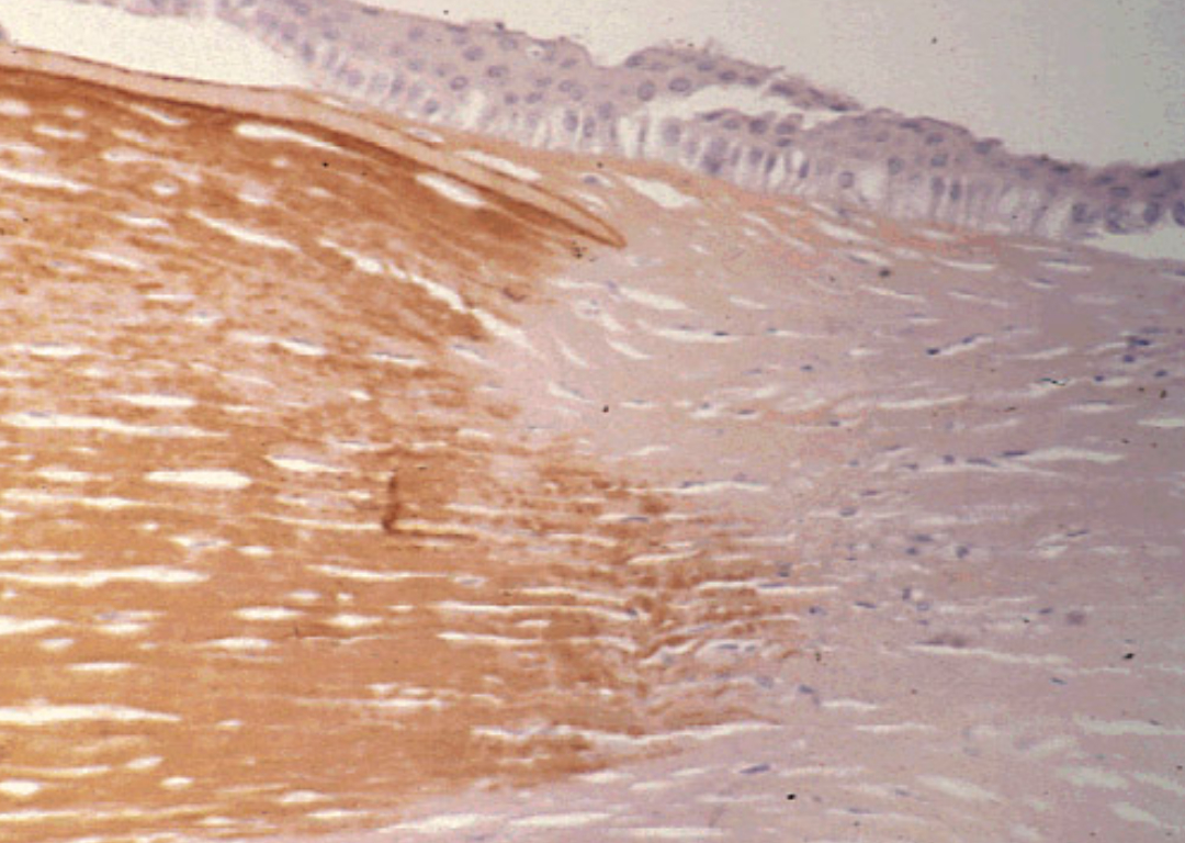
Photo credit: Klintworth GK, Wikimedia, CC BY 2.0