In a new study from LVPEI, Dr. Brijesh Takkar and others evaluated the safety and efficacy of fibrin glue in preventing vitreous hemorrhage after surgery for proliferative diabetic retinopathy.
Diabetic retinopathy is one of the consequences of diabetes mellitus, where high blood sugar slowly affects the retina, the innermost layer of the eye. When this condition reaches an advanced stage, known as proliferative diabetic retinopathy (PDR), it triggers an abnormal growth of fragile blood vessels in the retina that are prone to bleeding into the eye. PDR causes progressive vision loss and, if left untreated, will lead to blindness. The standard treatment is using lasers to control the abnormal blood vessels (laser photocoagulation), followed by vitrectomy surgery depending on severity of disease and visual deficit. However, surgery often fails as fragile blood vessels (bleeders) can continue to leak blood despite perfect surgery in up to a third of these eyes. Such postoperative vitreous cavity hemorrhage (POVCH) makes the vitreous murky and delays recovery. Stopping this bleeding requires an effective hemostat: an agent that blocks the bleeders at the back of the eye.
Fibrin glue is a biological adhesive made from two proteins involved in blood clotting, fibrinogen and thrombin. Fibrin glue is a well-known hemostat that can stop bleeding within 3 minutes of application. Moreover, it is non-toxic, biodegradable, and can be absorbed by the body. Fibrin glue has been shown to be effective at repairing retinal disorders such as macular holes or retinal tears. However, there are documented as well as anecdotal instances that indicate fibrin glue application can result in abnormal fibrous membrane growth. A randomized control trial—the gold standard of research—can help clarify fibrin glue’s safety and efficacy.
In a novel study published in the American Journal of Ophthalmology, Dr. Brijesh Takkar and others from LVPEI evaluated the efficacy and safety of fibrin glue in preventing early POVCH. Their single-masked, randomized controlled clinical trial, named ‘HaEmostasis with GLUe’ (HEGLU), included 20 patients with vitreous hemorrhage, half of whom were assigned as ‘cases,’ and the other half as ‘controls.’ Both groups underwent vitrectomy to remove the bloody vitreous, but only the cases received fibrin glue as a hemostat.
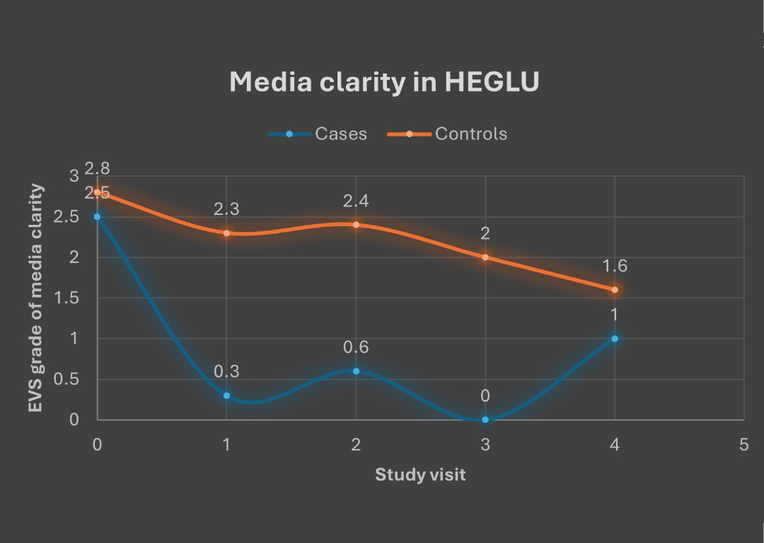
A week after surgery, cases had a much clearer vitreous (low vitreous haze) compared to the controls. This trend was re-checked and noted again at week 4. In fact, none of the cases had any vitreous haze at one month after surgery. Similarly, the vitreous cavity, the ocular space filled with vitreous, was observed to be optically clear in many more cases than in controls at both week 1 and week 4. Half of the control patients required secondary interventions for POVCH within 3 months of surgery. The cases had no such issue. While intermediate-term improvement in visual acuity was comparable for both groups, use of fibrin glue dramatically reduced the risk of recurrent bleeding into the vitreous without any side effects.
‘This is the first evidence stating the safety of fibrin glue application right over the extremely crucial and visually sensitive areas of the eye,’ remarks Dr. Brijesh Takkar, a consultant ophthalmologist at LVPEI and the primary author of this paper. ‘We found it safe in our clinical experiment, fortunately, also very useful in controlling retinal bleeding both during and after surgery for patients with PDR! The surgical technique is resource extensive, and easy to learn and adopt.’
Citation
Takkar, B., Ketkar, M. R., Narula, R., Tyagi, M., Dave, V. P., & Narayanan, R. (2025). A Pilot Randomized Controlled Trial Evaluating Hemostasis With Fibrin Glue During Surgery for Proliferative Diabetic Retinopathy. American Journal of Ophthalmology, 272, 117–125. Advance online publication. https://doi.org/10.1016/j.ajo.2025.01.016
Photo credit: Fig 1, Takkar et al.