In a new study, Drs. Vivek Singh, Sayan Basu, Vijay Kumar Singh, and others have developed a simple, precise, and reliable method for creating rabbit models with limbal stem cell deficiency (LSCD) disease using the Algerbrush II ophthalmic burr.
The cornea-the smooth, dome-shaped outer tissue on our eyes—is made of multiple layers of transparent epithelium, stacked together. A healthy cornea can repair its layers with stem cells in the limbus, a thin ring that separates the cornea from the sclera (the whites of the eye). Damage to the corneal epithelium, like in the case of calcium carbide induced ocular burns, can lead to the loss of limbal stem cells (LSCs). This loss cripples the cornea’s ability to regenerate, increasing the risk of corneal infections, clouding, and scarring. Such a permanent depletion of the limbus is called limbal stem cell deficiency (LSCD). While surgical means of remediation such as simple limbal epithelial transplantation (SLET) and cultivated limbal epithelial transplantation (CLET) remain staples, advancements in biomaterial research, like human tissue-engineered corneas, are next-generation LSCD treatments on the horizon. Before such experimental therapeutics can be applied to humans, it is crucial to first test their safety and efficacy on laboratory animals.
Animal models are thus pivotal for developing innovative diagnostics and novel therapies for LSCD management. In these models, LSCD is induced in laboratory strains of mice or rabbits either through surgery, chemicals (e.g., sodium hydroxide or mustard gas), or a combination of both. However, each of these options has limitations. Surgically induced LSCD requires the precision of a qualified surgeon, a luxury not feasible for many laboratories. In chemical injury models, it is challenging to limit the extent of corneal damage and resultant sequelae. The complications resulting from the imprecise depletion of LSCs can interfere with therapeutic research. What if there was a better way to create reliable LCSD animal models?
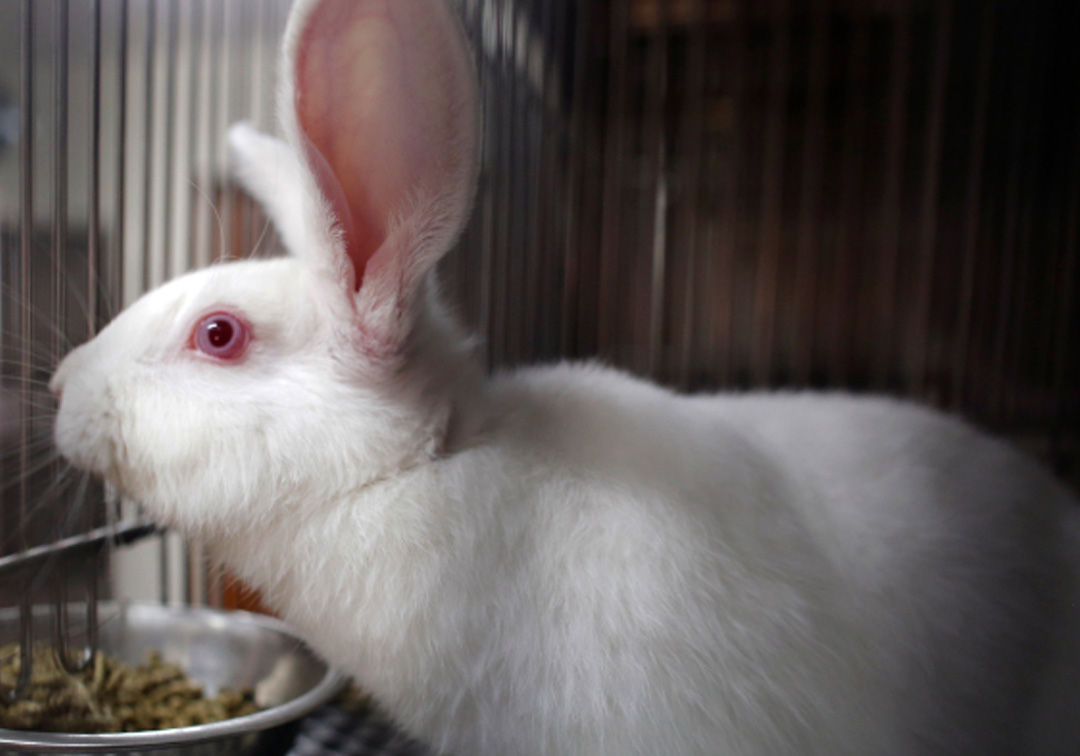
In a new study published in the journal Experimental Eye Research, Drs. Vivek Singh, Sayan Basu, Vijay Kumar Singh, and others from LVPEI used mechanical injury to generate LSCD in rabbit models. In this study, LSCD was induced using Algerbrush II, an ophthalmic burr used for grinding off foreign bodies from the ocular surface. Using a 1.0 mm rotating head, the team debrided the corneal and limbal epithelium of 18 male New Zealand White Rabbits that were 10- to 12-weeks-old. Among the 18 rabbits, 4 developed mild LSCD, while 10 had moderate, and 4 rabbits had severe LSCD. The LSCD was graded by the severity of parameters like corneal opacity, corneal thickness, and formation of new blood vessels. Defects in the corneal epithelium were assessed by staining the rabbit’s eye with fluorescein.
At the molecular level, the corneal epithelium is identified by expression of the protein K12 and LSCs are identified by the proteins p63 and ABCG2. Using qPCR, the team observed a significant decrease in the expression of these genes in rabbits with severe LSCD, confirming the loss of corneal epithelium and LSCs. ELISA testing of rabbit tears revealed an increase in the levels of inflammatory marker proteins (cytokines) like IL2 and IL17 and the wound healing marker MMP9. The authors believe that this mechanical injury model can reliably induce LSCD of various grades without requiring a surgeon’s expertise.
‘The present approach holds promise in establishing an efficient limbal stem cell deficiency model,’ says Dr. Vijay Kumar Singh, post-doctoral researcher at LVPEI and the first author of the paper. ‘This model can help us achieve a deeper understanding of ocular wound healing mechanisms and facilitate the exploration of innovative therapeutics for treating ocular surface defects.’
Citation
Singh VK, Kethiri AR, Pingali T, Sahoo A, Salman M, Koduri MA, et al. Development and validation of a reliable rabbit model of limbal stem cell deficiency by mechanical debridement using an ophthalmic burr. Exp Eye Res. 2023 Sep 26;236:109667.
Photo credit: Laboratory Rabbits; by Otwarte Klatki; CC BY 2.0.