Cornea and Anterior Segment
The Cornea and Anterior Segment unit at LVPEI has treated patients from all over India and the neighboring countries. It specializes in the treatment of the entire range of corneal disorders and infections. This unit has set a record of sorts by performing more than 1000 corneal transplants in a single calendar year (2003), using corneas harvested by LVPEI's Eye Bank. The Eye Bank also distributes corneas to hospitals in Hyderabad and other places.
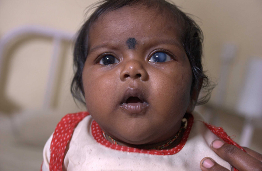
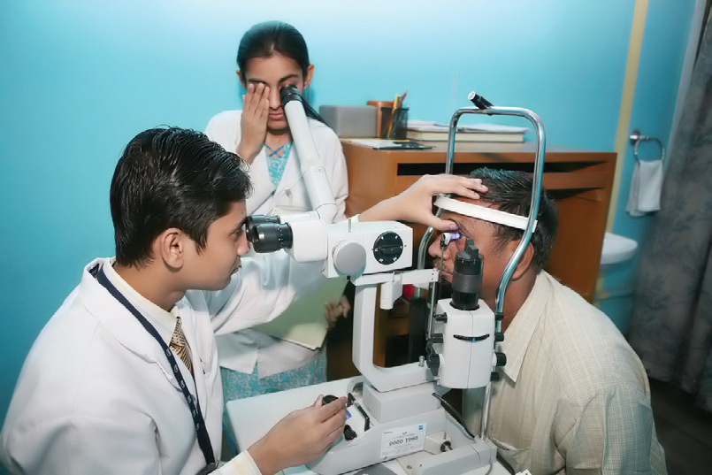
Apart from cataract surgery and corneal transplants, clinicians in this service handle a range of other ocular complications, from infections on the surface of the eye and eyelids to trauma care. Clinicians also work closely with scientists at the institute to develop new and more cost-effective treatments. Among the several research projects at the Institute is the stem cell research program, which has made possible an innovative treatment procedure for persons with ocular surface damage.
Ophthalmologists at LVPEI have expertise and experience in laser assisted surgical procedures. The Institute has been providing laser assisted refractive surgeries for many years with extremely successful results. Recently the Wavefront Assisted Customized Vision Correction Technology was added to the list of corneal services. This new equipment from Bausch & Lomb enables patients to benefit from the latest advancements in refractive eye surgery.
What is a Corneal Transplant?
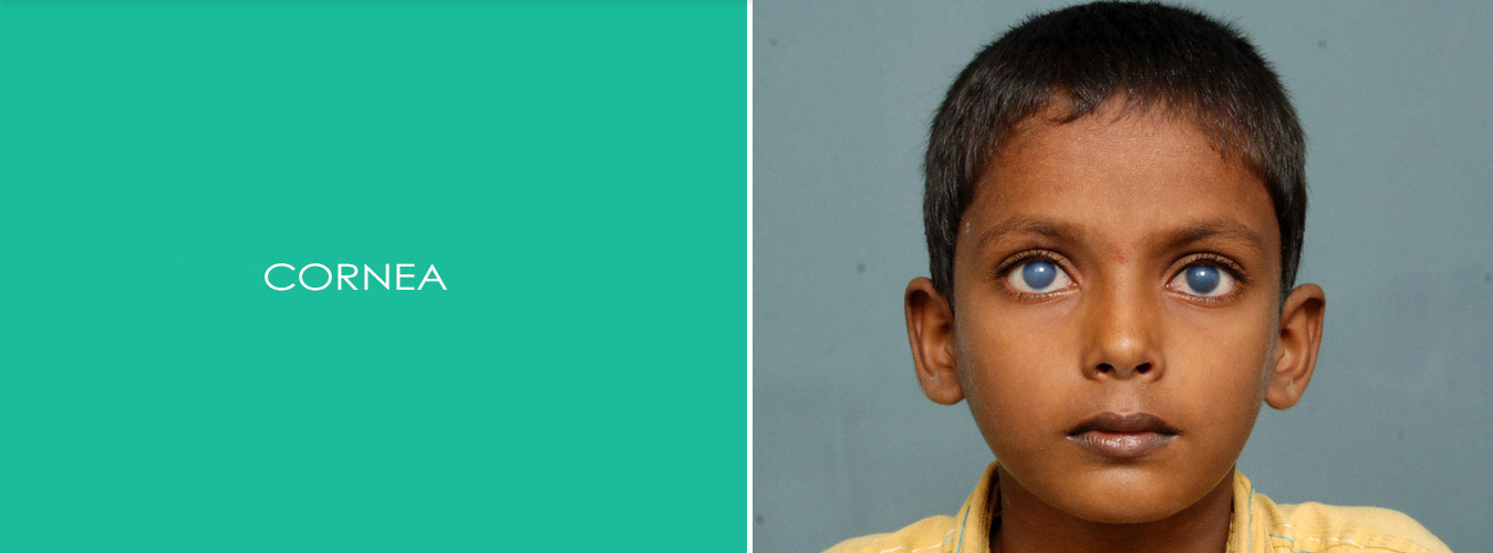
A transplant is the replacement of damaged or diseased tissues or organs with healthy tissues or organs. In a corneal transplant, the cloudy or warped cornea is replaced with a healthy cornea. If the new cornea heals without problems, there may be tremendous improvement in vision.
The healthy corneal tissue used for transplantation is supplied by an Eye Bank. Eye Banks work round the clock to collect, evaluate, and store donated corneas. The corneas are collected from human donors within hours of death. Stringent tests are done to ensure the safety of the person receiving the cornea. The Eye Bank verifies the donor's medical history and cause of death, and performs blood tests to ensure that the deceased person did not have any contagious disease, such as AIDS or hepatitis.
Since the cornea was one of the first parts of the body to be transplanted, corneal transplants remain one of the most common, and most successful, of all transplants.
Anything you see is an image that enters your eye in the form of light. The different parts of your eye collect this light and send a message to your brain, enabling you to see. For perfect vision all the parts of your eye need to work properly.
- The cornea is the clear, outer layer of the eye.
- The pupil is an opening that lets light enter the eye.
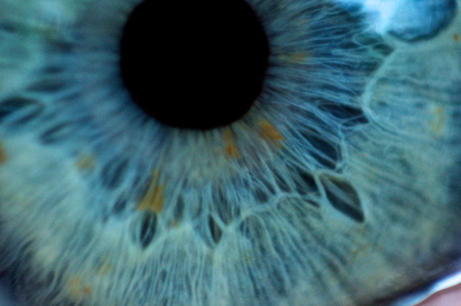
- The iris, the colored part of the eye, makes the pupil larger or smaller.
- The lens bends to focus light onto the retina.
- The retina receives light that has been focused by the cornea and lens.
- A clear (vitreous) gel fills the inside of the eye, giving it shape.
The cornea is clear to let light into the eye, and curved to focus the light rays.
